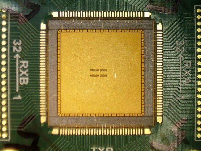
The Massachusetts Institute of Technology has introduced a compact ultrasound device for monitoring breast tissue. The device weighs 520 grams and can produce images of tissue at depths of more than 11 centimeters — on par with clinical systems, but with much lower power consumption.
The development is aimed primarily at women with dense breast tissue, nearly 40% of whom often face difficulties detecting changes on mammograms. Instead of a large panel with thousands of sensors, the device uses a compact housing with 128 sensors of a special geometry. By distributing the transmitting and receiving elements, the system creates a detailed 3D image comparable to systems with eight times more components.
A new signal-processing method has also been applied — chirps, or ultrasound that changes frequency smoothly. It penetrates tissue better and makes it possible to use simpler electronics without overheating.
For now, the device has been tested only on a limited basis and requires further clinical research. Still, the fact that clinical-quality imaging was achieved with minimal power changes expectations about what portable systems can do. If the technology passes large-scale clinical trials and receives regulatory approval, home monitoring between routine checkups could become a reality.