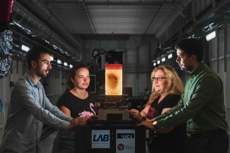
Specialists from University College London and the European Synchrotron Radiation Facility have presented an open online atlas of human organs, already being called a “Google Maps” for the body. The new resource lets users literally “travel” inside organs — from an overall view of the heart or lungs down to the level of individual muscle fibers.
The project’s main innovation is its combination of high precision and full accessibility. The atlas runs directly in a browser and does not require special equipment. It includes 298 three-dimensional image sets and covers 10 organ types. The database contains both healthy tissue and tissue affected by disease. The key technology is hierarchical phase-contrast tomography (HiP-CT). It makes it possible to scan an entire organ while also capturing high detail without destroying it.
The project has broad practical value. It is changing the approach to medical education. Students gain access not to diagrams, but to real organs: they can rotate them, “dive” into the tissue and study the structure in its natural complexity. This makes learning more visual and closer to practice. In addition, the atlas is becoming a valuable source of data for training artificial intelligence systems.